Leg Bone Diagram : Knee Human Anatomy Function Parts Conditions Treatments
Leg Bone Diagram : Knee Human Anatomy Function Parts Conditions Treatments. There are two types of cartilaginous joints: Muscle anatomy diagram 12 photos of the muscle anatomy diagram canine muscle anatomy diagram, dog muscle anatomy diagram, lower leg muscle anatomy diagram, muscle anatomy of human back, tricep muscle anatomy diagram, human muscles, canine muscle anatomy diagram, dog muscle anatomy diagram, lower leg muscle anatomy. Related posts of muscles and tendons of the leg muscle anatomy diagram. To explain the term in layman's language, it is the heel bone in the skeletal system. The muscles of the leg is a big topic, so take your time to learn it fully.
ads/bitcoin1.txt
Compact bone is the denser, stronger of the two types of bone tissue ( (figure) ). The knee is one of the largest and most complex joints in the body. If you enjoyed learning the muscles of the leg with our quizzes and labeling exercises, look no further than our library of free quiz guides on tricky exam topics like the cranial nerves, bones of the skull and reproductive systems. Next to the tibia is the fibula, the thinner, weaker bone of the lower leg. Pelvic bone labeled 12 photos of the pelvic bone labeled pelvic bone labeled, pelvic bone labeling quiz.
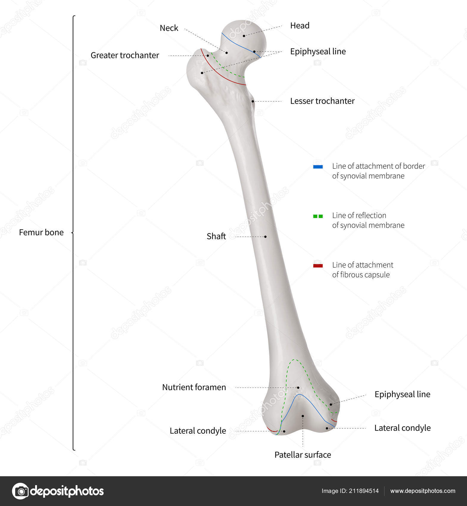
Infographic Diagram Human Femur Bone Leg Bone Anatomy System Anterior Stock Photo Image By C K Intarapong Gmail Com 211894514 from st4.depositphotos.com The knee joint is the largest joint in the body and is primarily a hinge joint, although some sliding and rotation occur. As a result, it doesn't play any crucial role in weight bearing. The knee joint is the largest joint in the body and is primarily a hinge joint, although some sliding and rotation occur. If you enjoyed learning the muscles of the leg with our quizzes and labeling exercises, look no further than our library of free quiz guides on tricky exam topics like the cranial nerves, bones of the skull and reproductive systems. The foot bones shown in this diagram are the talus, navicular, cuneiform, cuboid, metatarsals and calcaneus. Related posts of diagram of leg bones pelvic bone labeled. It can be found under the periosteum and in the diaphyses of long bones, where it provides support and protection. Educational diagram with pronated, normal and supinated compared examples with bone titles.
Compact bone is the denser, stronger of the two types of bone tissue ( (figure) ).
ads/bitcoin2.txt
The tarsal bones in the foot are located amongst tibia, metatarsal bones, and fibula. The hip itself is a ball and socket joint, much like the shoulder.the structures necessary to create this joint are the socket, the joint capsule, muscle, ligaments, and the neck. Related posts of muscles and tendons of the leg muscle anatomy diagram. If you enjoyed learning the muscles of the leg with our quizzes and labeling exercises, look no further than our library of free quiz guides on tricky exam topics like the cranial nerves, bones of the skull and reproductive systems. Some common causes of leg pain include: The fibula is smaller, thinner, and laterally positioned compared to the tibia. Leg pain can also be caused by blood clots, varicose veins or poor circulation. In this image, you will find femur, medial epicondyle of the femur, patella, tibial tuberosity, anterior rest of the tibia, a medial surface of the tibia, lateral epicondyle of the femur, head of the fibula, fibula, medial malleolus of the tibia, lateral. Pelvic bone labeled 12 photos of the pelvic bone labeled pelvic bone labeled, pelvic bone labeling quiz. Also called the shin bone, the tibia is the longer of the two bones in the. It can be found under the periosteum and in the diaphyses of long bones, where it provides support and protection. Related posts of diagram of leg bones pelvic bone labeled. In this anatomy lesson, i'm going to cover the anatomy of the tibia and fibula bones of the anatomical leg, which is the section between the knee and ankle.
Beside that, we also come with more related ideas as follows free printable human anatomy coloring pages, lower leg muscle diagram blank and lower limb bones unlabeled. With different grades of sprains depending on severity. Original file at image/png format. The bones together make up the hip. The knee joint is the largest joint in the body and is primarily a hinge joint, although some sliding and rotation occur.
Bones Of The Human Leg 17 Download Scientific Diagram from www.researchgate.net Related posts of muscles and tendons of the leg muscle anatomy diagram. It is also known as the calf bone, as it. Leg pain can also be caused by blood clots, varicose veins or poor circulation. Related posts of diagram of leg bones pelvic bone labeled. Tibia and fibula bone anatomy. This area is commonly referred to as the calf. The foot bones shown in this diagram are the talus, navicular, cuneiform, cuboid, metatarsals and calcaneus. 10 / 10 ( 1 vote ) leg bone anatomy diagram diagram of human leg human anatomy diagram.
At the same time, the bones and joints of the leg and foot must be strong enough to support the body's weight while remaining.
ads/bitcoin2.txt
Tibia and fibula bone anatomy. Some common causes of leg pain include: As a result, it doesn't play any crucial role in weight bearing. The smaller bone that runs alongside the tibia (fibula) and the. The pubis, ischium, and ilium together constitute the pelvis while the thigh bone is the femur. Our goal is that these leg anatomy worksheets pictures gallery can be a direction for you, bring you more references and also make you have a great day. Leg pain can also be caused by blood clots, varicose veins or poor circulation. Most leg pain results from wear and tear, overuse, or injuries in joints or bones or in muscles, ligaments, tendons or other soft tissues. The fibula is smaller, thinner, and laterally positioned compared to the tibia. In this image, you will find femur, medial epicondyle of the femur, patella, tibial tuberosity, anterior rest of the tibia, a medial surface of the tibia, lateral epicondyle of the femur, head of the fibula, fibula, medial malleolus of the tibia, lateral. The knee joint is the largest joint in the body and is primarily a hinge joint, although some sliding and rotation occur. The muscles of the leg is a big topic, so take your time to learn it fully. Educational diagram with pronated, normal and supinated compared examples with bone titles.
In this image, you will find femur, medial epicondyle of the femur, patella, tibial tuberosity, anterior rest of the tibia, a medial surface of the tibia, lateral epicondyle of the femur, head of the fibula, fibula, medial malleolus of the tibia, lateral. Some types of leg pain can be traced to problems in your lower spine. Our goal is that these leg anatomy worksheets pictures gallery can be a direction for you, bring you more references and also make you have a great day. The major bones of the leg are the femur (thigh bone), tibia (shin bone), and adjacent fibula, and these are all long bones.the patella (kneecap) is the sesamoid bone in front of the knee.most of the leg skeleton has bony prominences and margins that can be palpated and some serve as anatomical landmarks that define the extent of the leg. The tibia and the fibula, at the top of the ankle joint.
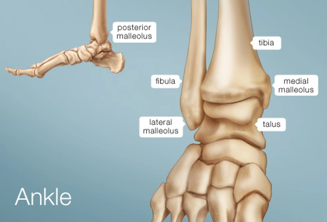
Ankle Human Anatomy Image Function Conditions More from img.webmd.com These muscles work together to produce movements such as standing, walking, running, and jumping. Original file at image/png format. The lower leg is comprised of two bones, the tibia and the smaller fibula. 10 / 10 ( 1 vote ) leg bone anatomy diagram diagram of human leg human anatomy diagram. Compact bone is the denser, stronger of the two types of bone tissue ( (figure) ). The thigh bone, or femur, is the large upper leg bone that connects the lower leg bones (knee joint) to the pelvic bone (hip joint). At the same time, the bones and joints of the leg and foot must be strong enough to support the body's weight while remaining. The bones together make up the hip.
Original file at image/png format.
ads/bitcoin2.txt
These landmarks are the anterior superior iliac spine. The fibula is smaller, thinner, and laterally positioned compared to the tibia. The knee is one of the largest and most complex joints in the body. The femur, or thigh bone, is the single bone of the thigh region (figure 6.51). Original file at image/png format. Related posts of muscles and tendons of the leg muscle anatomy diagram. It can be found under the periosteum and in the diaphyses of long bones, where it provides support and protection. The foot bones shown in this diagram are the talus, navicular, cuneiform, cuboid, metatarsals and calcaneus. If you enjoyed learning the muscles of the leg with our quizzes and labeling exercises, look no further than our library of free quiz guides on tricky exam topics like the cranial nerves, bones of the skull and reproductive systems. In this image, you will find femur, medial epicondyle of the femur, patella, tibial tuberosity, anterior rest of the tibia, a medial surface of the tibia, lateral epicondyle of the femur, head of the fibula, fibula, medial malleolus of the tibia, lateral. The lower leg is comprised of two bones, the tibia and the smaller fibula. The pubis, ischium, and ilium together constitute the pelvis while the thigh bone is the femur. The knee joins the thigh bone (femur) to the shin bone (tibia).
ads/bitcoin3.txt
ads/bitcoin4.txt
ads/bitcoin5.txt
0 Response to "Leg Bone Diagram : Knee Human Anatomy Function Parts Conditions Treatments"


0 Response to "Leg Bone Diagram : Knee Human Anatomy Function Parts Conditions Treatments"
Post a Comment